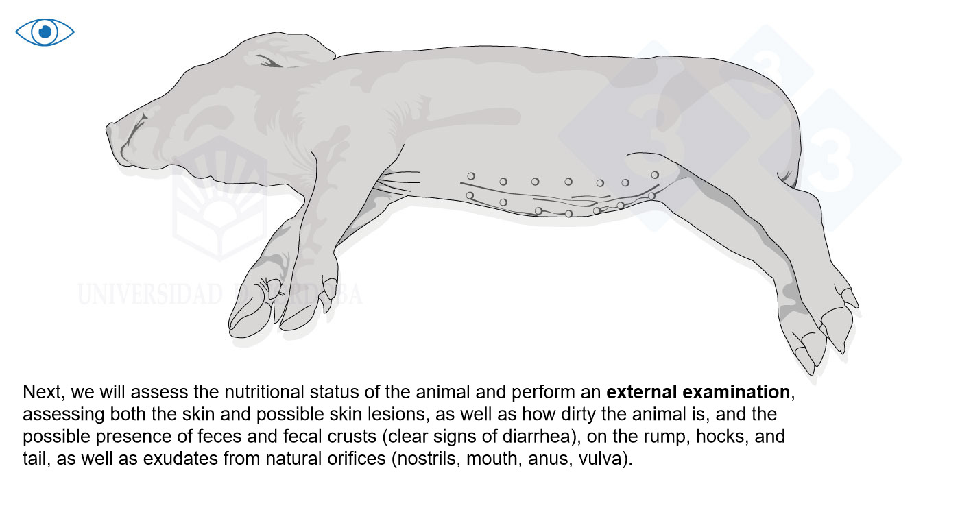
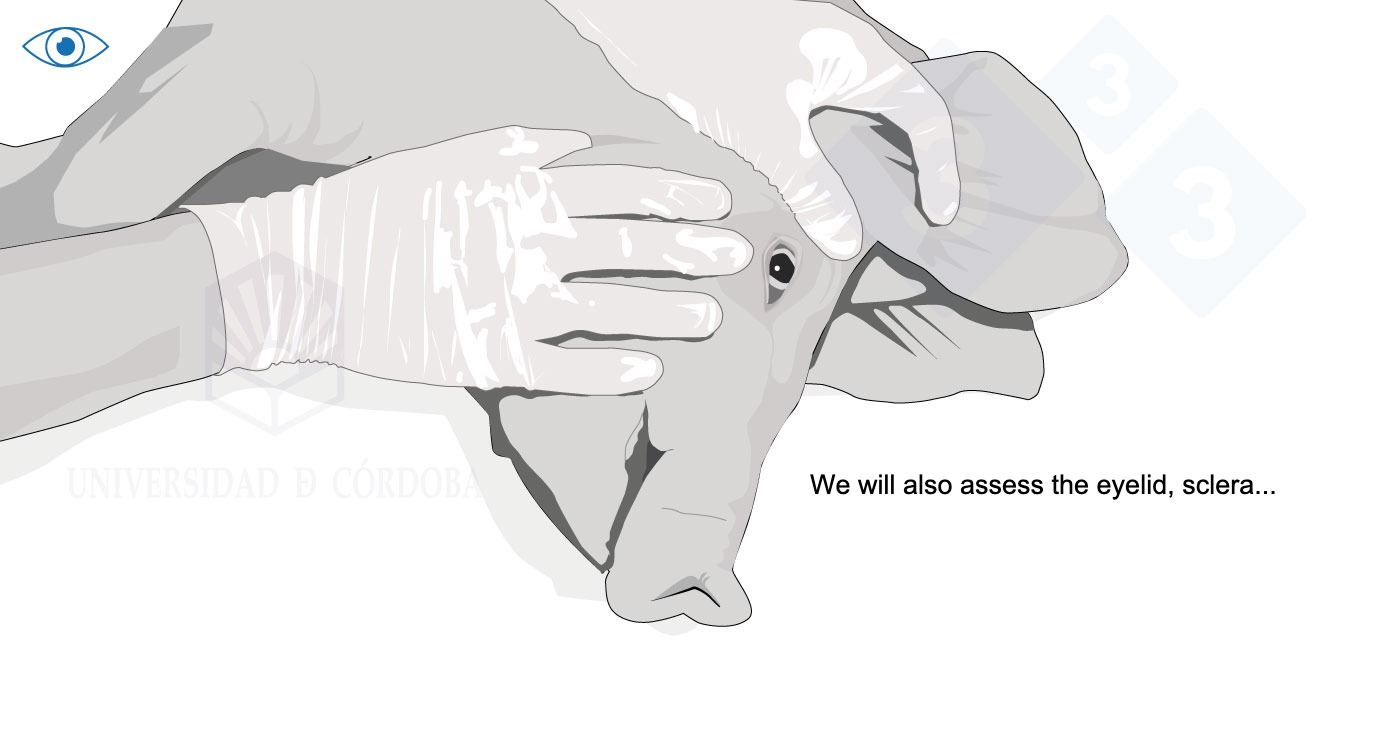
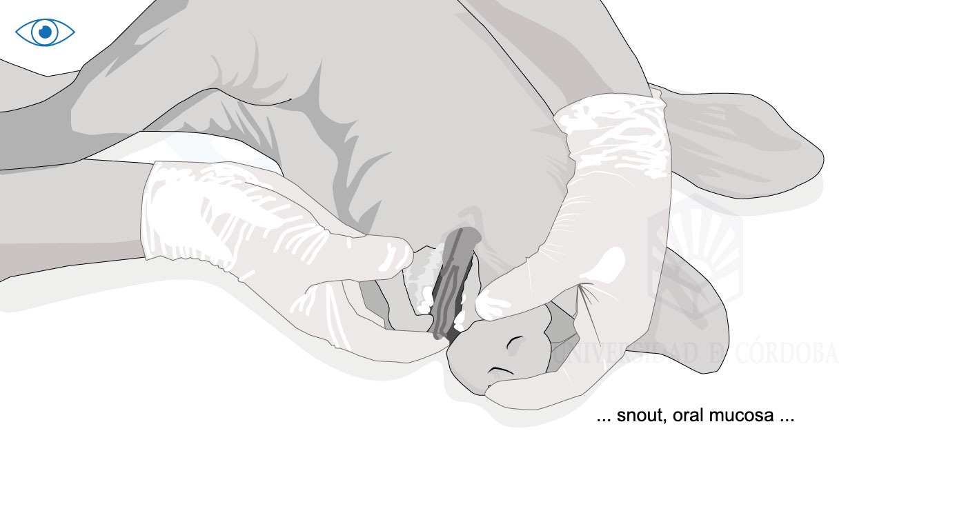
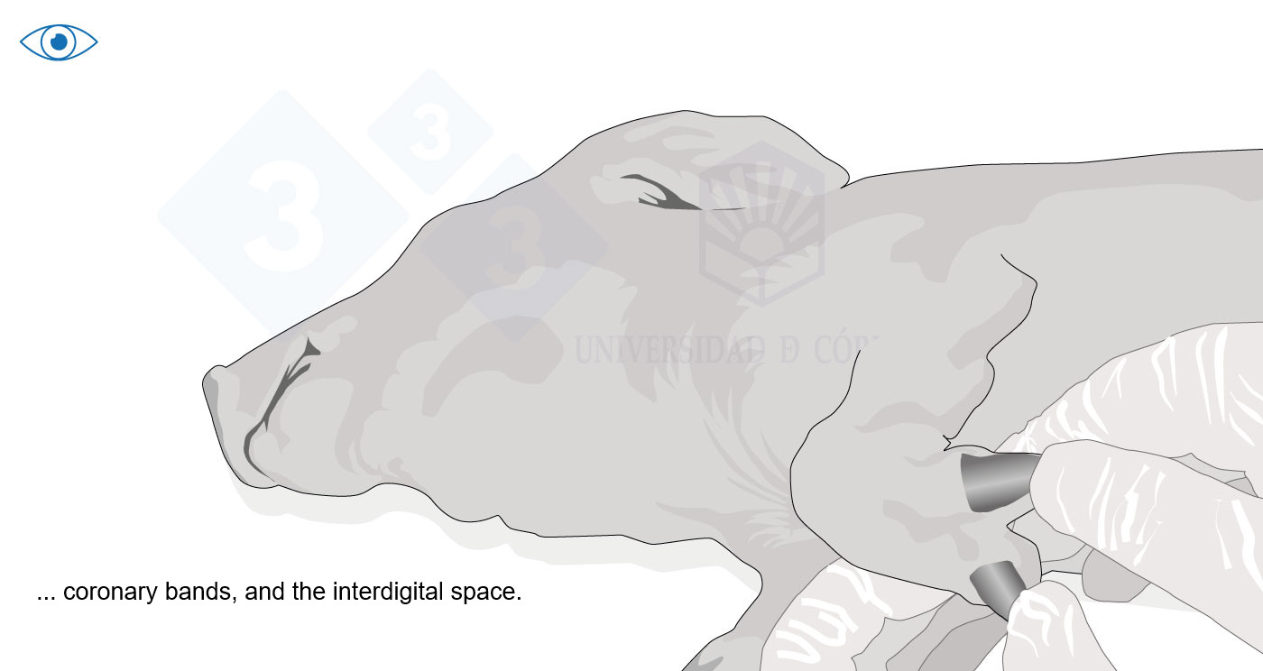
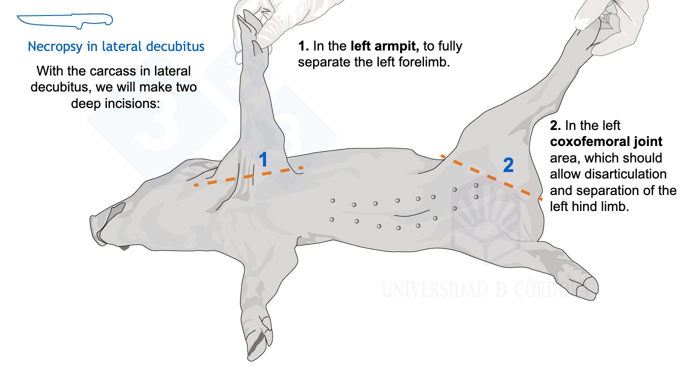
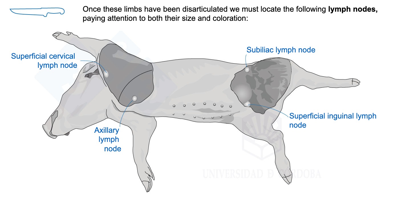
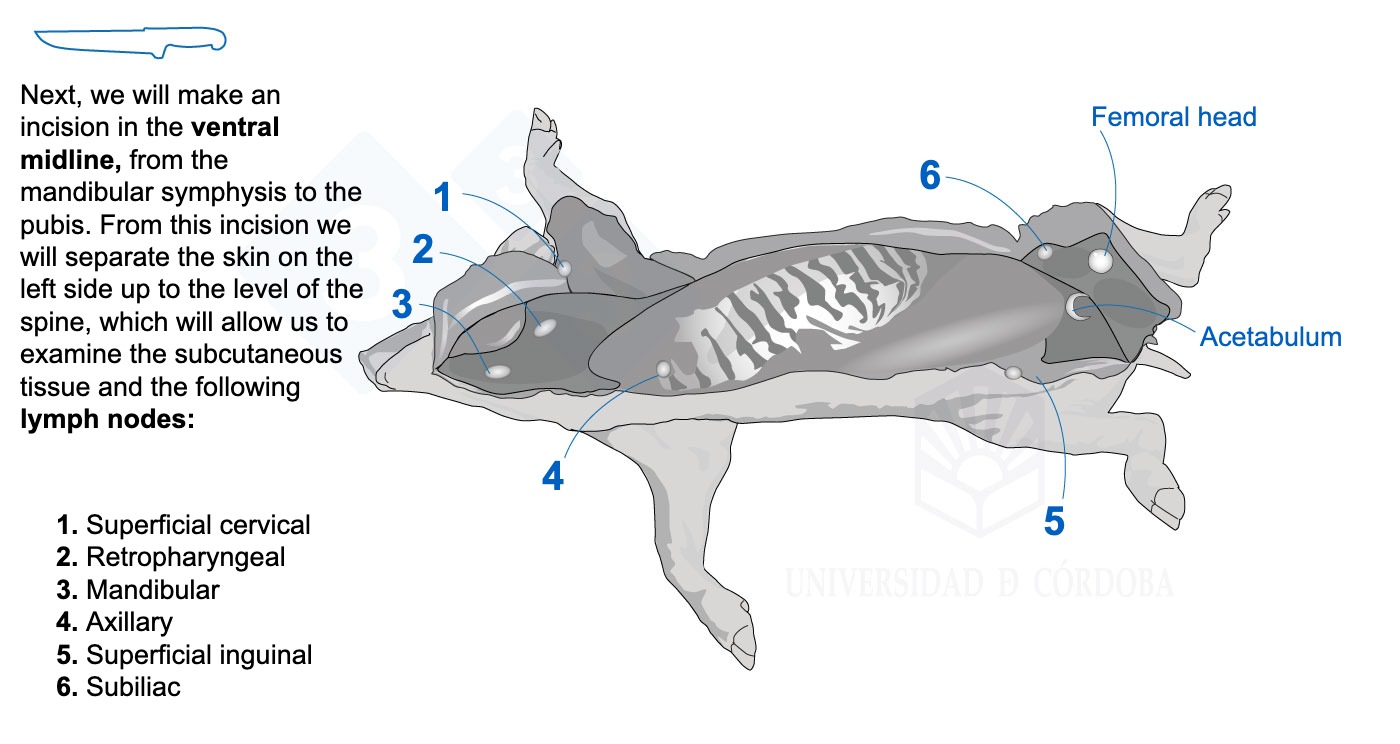
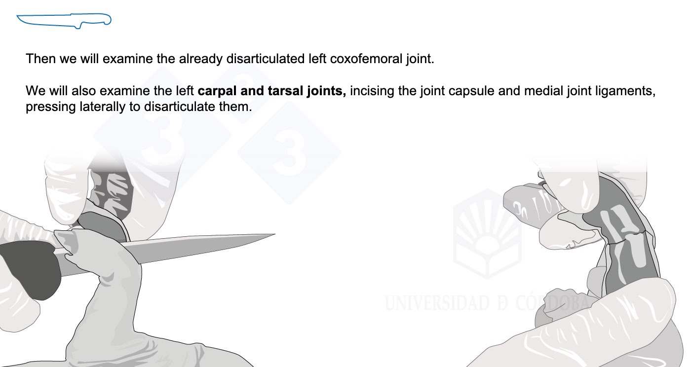
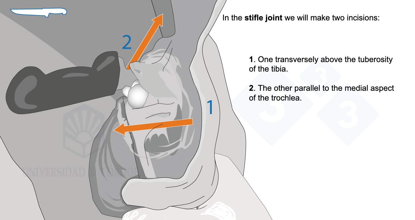
We will start with the necropsy of a piglet in lateral decubitus. In this first installment, we will do the external examination, separate the left forelimb and hind limb, and make a ventral midline incision, which will allow us to assess the lymph nodes and subcutaneous tissue.
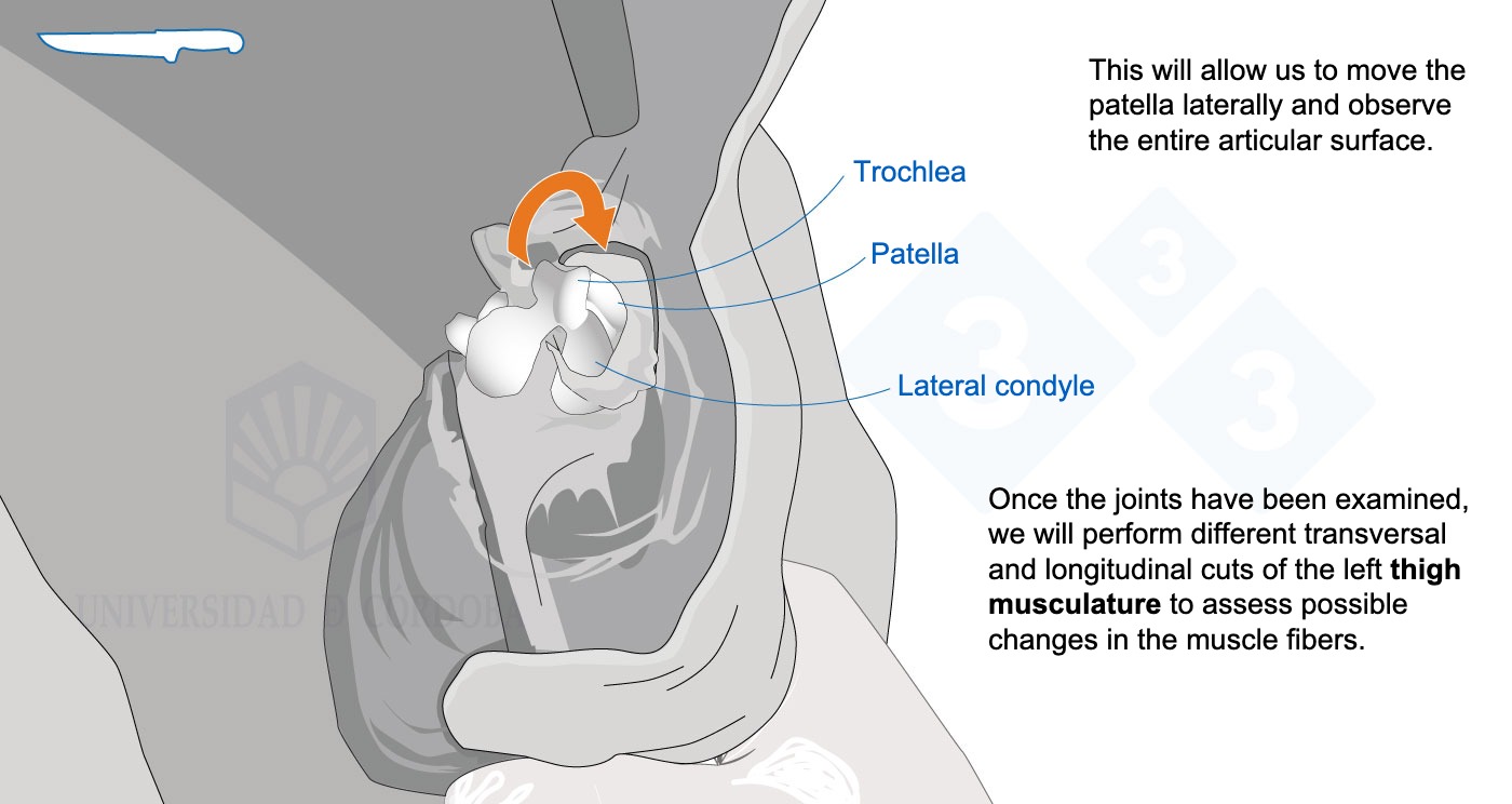
We will also assess the coxofemoral, carpal, tarsal, and femorotibial joints.

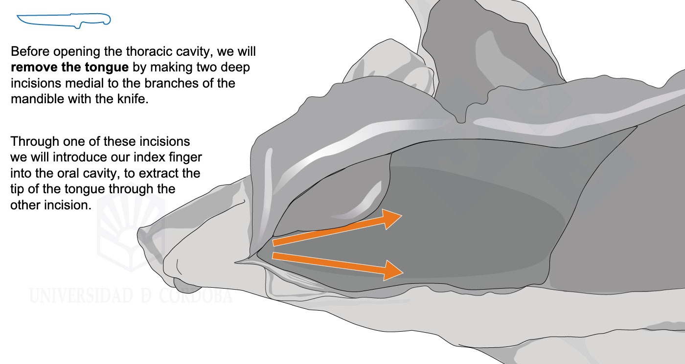
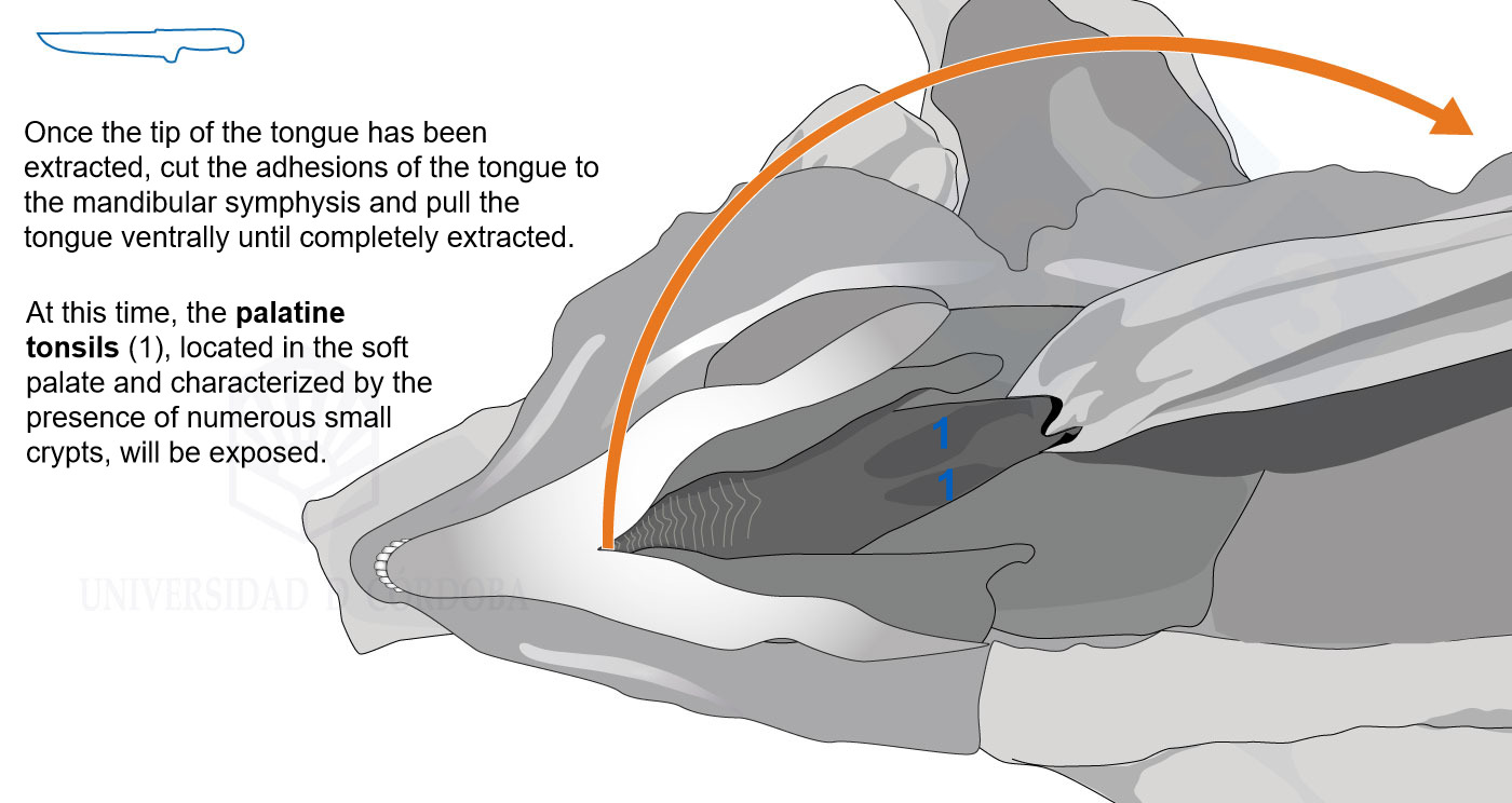
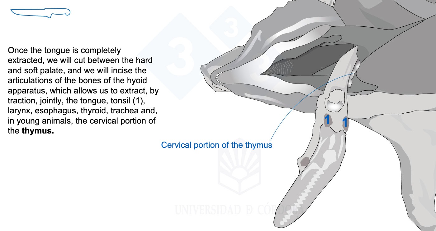
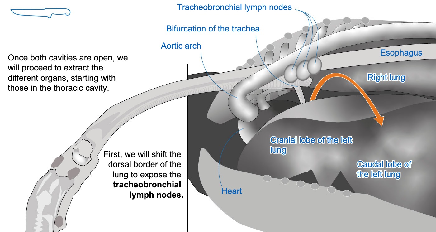
We will extract the tongue, together with the esophagus and the trachea, which will expose the palatine tonsils and the thymus.
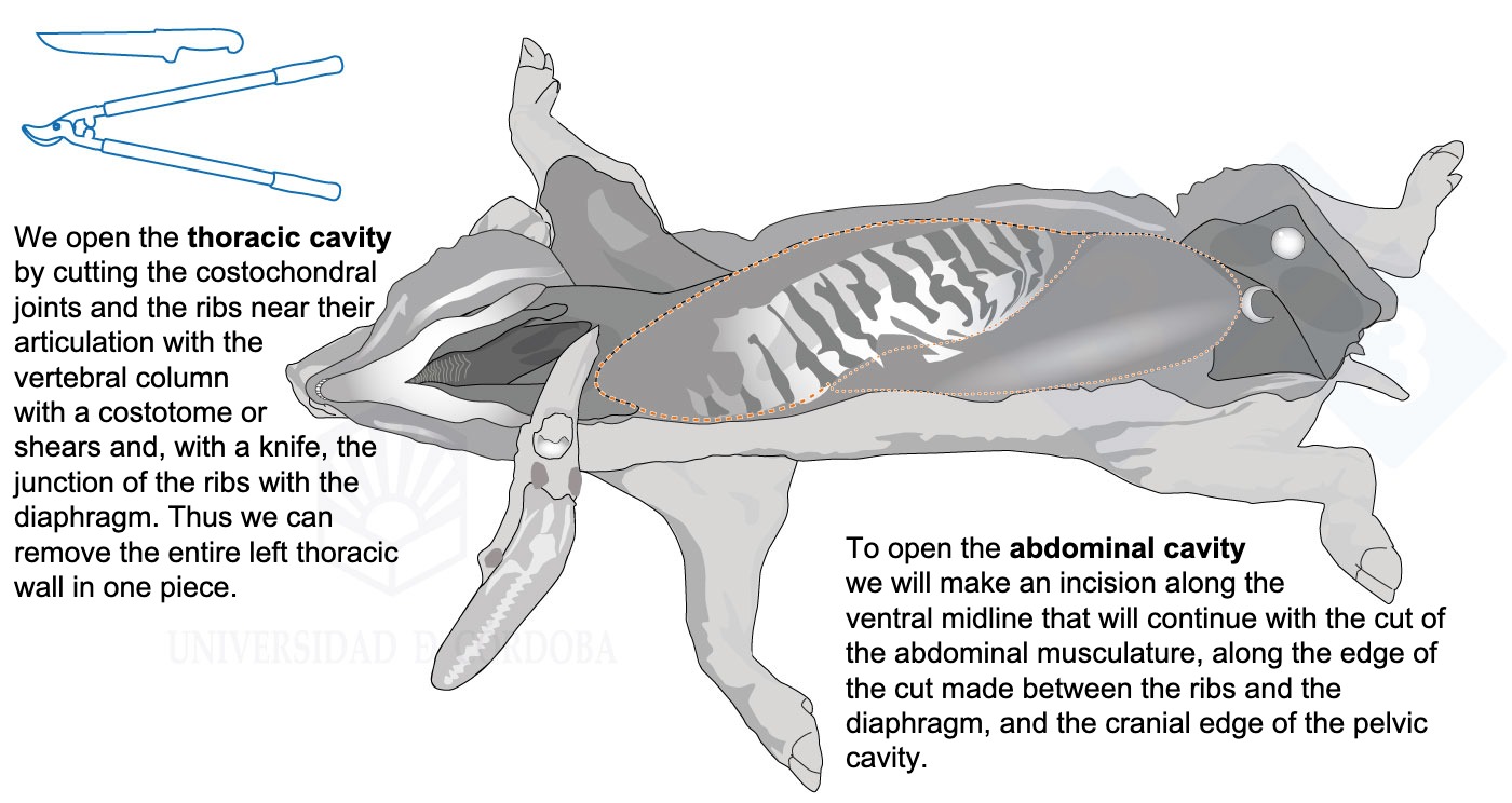
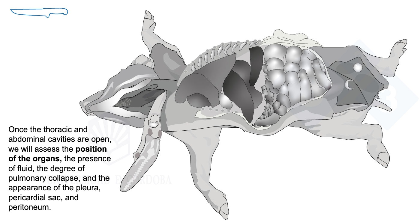
Finally, we will open the thoracic and abdominal cavity, allowing us to observe the degree of pulmonary collapse and the appearance of the pleurae, the pericardial sac, and the peritoneum.
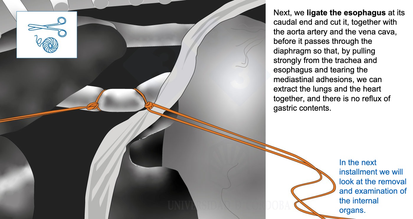
Finally, we'll ligate the esophagus at its caudal end.
In the next installment, we will look at the removal and examination of the internal organs.
Click on any part of the image to advance. Use the left arrow to go backward. Use the square icon at the bottom right to view the presentation in full screen.


